Overview
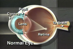
The cornea is the clear “window” in the front of the eye that allows light rays to enter.
The cornea has three layers – the outer epithelium (or skin), a middle area called stroma and a delicate, single-celled inner lining called the endothelium. The endothelium acts as a barrier to prevent water inside the eyeball from moving into and swelling the other layers of the cornea. The cells of the endothelium actively pump water from the cornea back into the eye.

If the endothelium does not function normally, then water moves into the cornea causing swelling. Swelling causes clouding of the cornea and blurred vision. The more corneal swelling or “edema,” the more severely the vision is blurred. Eventually, the outer corneal layer (epithelium) also takes on water, resulting in pain and more severe vision impairment. Epithelial swelling reduces vision by changing the normal curvature of the cornea. It causes a sight-limiting haze to develop. Epithelial swelling may also form small “blisters” on the corneal surface. When these “blisters” burst, extreme pain can occur.
Endothelial cells can be counted with special photographic methods. Most people are born with approximately 4,000 cells per square millimeter of the endothelial surface. These cells do not divide and cannot reproduce or replace themselves. As we age, we gradually lose endothelial cells.
